Galerie obrázků
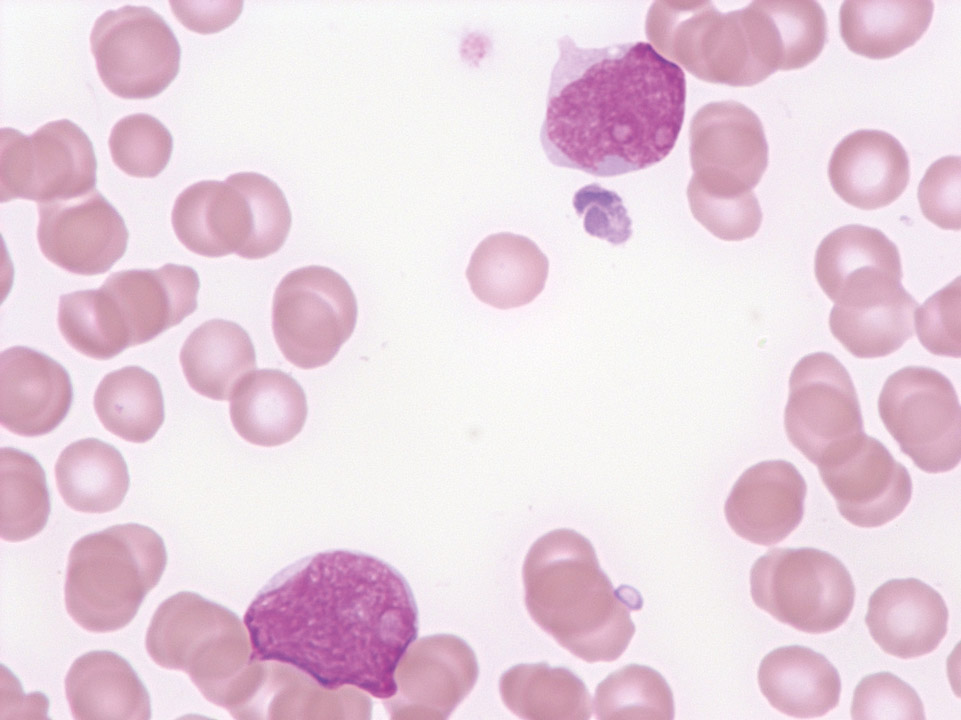
It is extremely rare that cancerous cells leak into the peripheral blood. Here, cells from a breast carcinoma are visible on the blood film.
<p>It is extremely rare that cancerous cells leak into the peripheral blood. Here, cells from a breast carcinoma are visible on the blood film.</p>
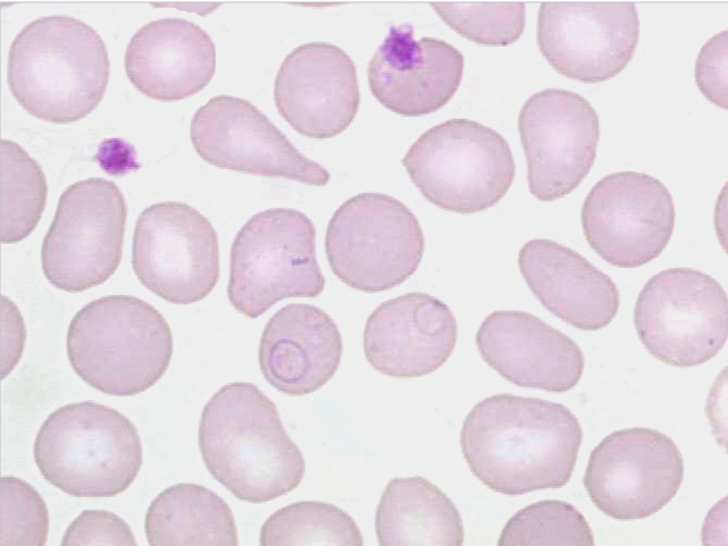
Cabot rings are round to oval or loop-like, fine, red-purple inclusions in red blood cells. They are remnants of the mitotic spindle apparatus. They can be seen with severe anaemia, e.g. thalassaemia, but are unspecific.
<p>Cabot rings are round to oval or loop-like, fine, red-purple inclusions in red blood cells. They are remnants of the mitotic spindle apparatus. They can be seen with severe anaemia, e.g. thalassaemia, but are unspecific. </p>
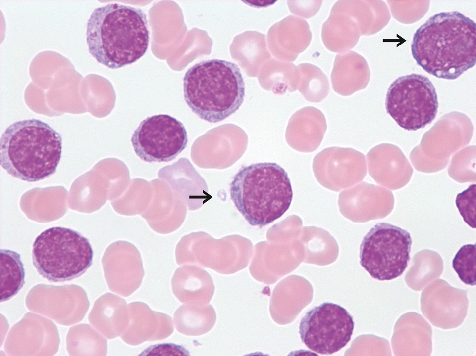
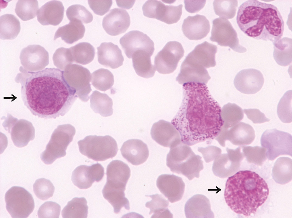
Transitional form of chronic lymphocytic leukaemia/prolymphocytic leukaemia with a white blood cell count of 250,000/μL. Prolymphocytes are indicated (->).
<p>Transitional form of chronic lymphocytic leukaemia/prolymphocytic leukaemia with a white blood cell count of 250,000/μL. Prolymphocytes are indicated (->).</p>
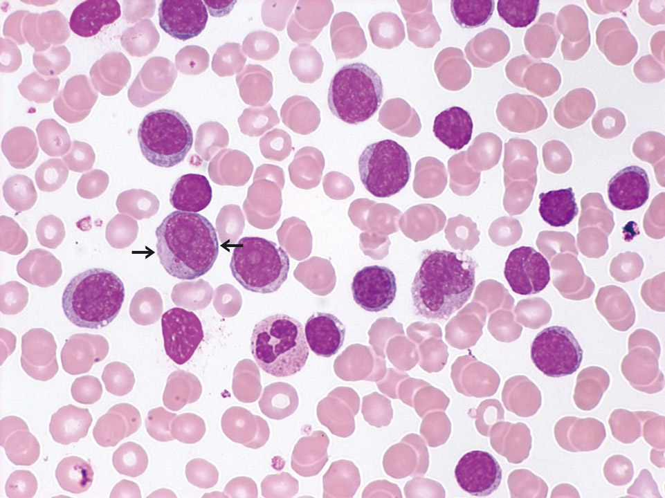
Chronic lymphocytic leukaemia with numerous prolymphocytes (->) (CLL/PL) in the peripheral blood (May-Grünwald-Giemsa stain).
<p>Chronic lymphocytic leukaemia with numerous prolymphocytes (->) (CLL/PL) in the peripheral blood (May-Grünwald-Giemsa stain). </p>
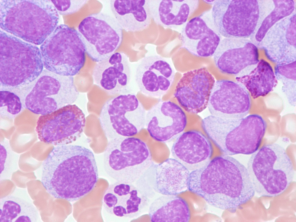
Blood film of a patient with chronic myelogenous leukaemia (CML). The extremely high white blood cell count of 680,000/μL caused a hyperviscosity syndrome and led to total and only partially reversible deafness.
<p>Blood film of a patient with chronic myelogenous leukaemia (CML). The extremely high white blood cell count of 680,000/μL caused a hyperviscosity syndrome and led to total and only partially reversible deafness. </p>
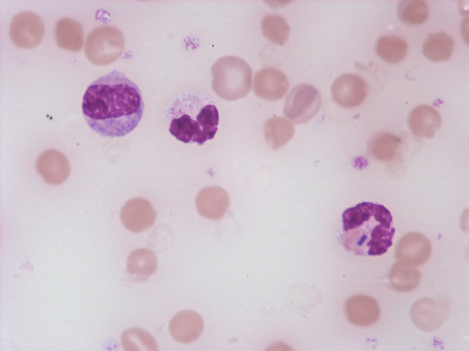
The barrel-shaped bacillus visible in the right granulocyte is Clostridium perfringens from a case of septic gangrene. (The reddish background is caused by massive red blood cell lysis. The white blood cells are about to dissolve as well.)
<p>The barrel-shaped bacillus visible in the right granulocyte is Clostridium perfringens from a case of septic gangrene. (The reddish background is caused by massive red blood cell lysis. The white blood cells are about to dissolve as well.)</p>
